HOW IT WORKS · EXAMPLE
How to read your results
The diagram and scenario below are illustrative. They are not a real person’s analysis or a before-and-after result, and will not be saved to your history.
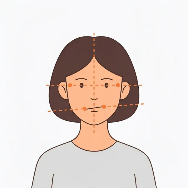
1. What was observed in the photo
For example, “a difference in mouth-corner height was observed” describes the relative positions of two points in the photo. It does not identify a weak muscle or tell you which side to chew on.
2. Check capture conditions first
Head tilt, expression, and an obscured face make comparison harder. If a result differs, check again with the same camera, distance, lighting, and a relaxed expression. Estimates can still vary under similar conditions.
3. The score is a custom observation index
A higher overall score means a smaller calculated left-right difference. It is not a clinical measure of normality, beauty, or treatment need. In contrast, a higher area deviation index means a larger observed difference.
4. Record and choose a next step
Use history to review observations alongside capture conditions. Recommended corrective exercises are general self-care information, not a medical prescription. Pain or new movement problems call for professional assessment rather than a self-check.